Fotosearch Royalty Free Stock Photography
Digital images licensed by Publitek, Inc.
Fotosearch and Photosearch are trademarks of Fotosearch, LLC
All rights reserved © 2026-01-25
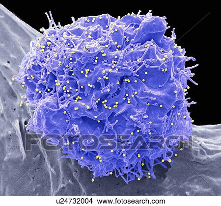
HIV infected cell. Coloured scanning electron micrograph (SEM) of a 293T cell infected with the human immunodeficiency virus (HIV, yellow dots). Small spherical virus particles, visible on the surface, are in the process of budding from the cell... Picture

HIV infected cell. Coloured scanning electron micrograph (SEM) of a 293T cell infected with the human immunodeficiency virus (HIV, yellow dots). Small spherical virus particles, visible on the surface, are in the process of budding from the cell...
u24732004 | Lushpix | Royalty Free
Add to Lightbox
Share Image
Keywords
293t, aids, antiviral, cell, cell-culture, cellular, colored, coloured, defence, defense, deoxyribonucleic-acid, dna, embryonic-kidney, exosome, false-coloured, gene-therapy, hiv, infection, medical, medicine, protein, research, scanning-electron-micrograph, scanning-electron-microscopy, sem, therapeutic, viral, virus, stock image, images, royalty free photo, stock photos, stock photograph, stock photographs, picture, pictures, graphic, graphics, fine art prints, print, poster, posters, mural, wall murals, u24732004
293t, aids, antiviral, cell, cell-culture, cellular, colored, coloured, defence, defense, deoxyribonucleic-acid, dna, embryonic-kidney, exosome, false-coloured, gene-therapy, hiv, infection, medical, medicine, protein, research, scanning-electron-micrograph, scanning-electron-microscopy, sem, therapeutic, viral, virus, stock image, images, royalty free photo, stock photos, stock photograph, stock photographs, picture, pictures, graphic, graphics, fine art prints, print, poster, posters, mural, wall murals, u24732004
Show Keywords
- Mobile/Small Web Resolution (150 KB)3.4" x 3" @ 72dpi JPG13
- Web Resolution (500 KB)6.2" x 5.5" @ 72dpi JPG32
- Low Resolution (1 MB)8.7" x 7.8" @ 72dpi JPG68
- Medium Resolution (10 MB)6.6" x 5.9" @ 300dpi JPG140
- High Resolution (28 MB)11.1" x 9.8" @ 300dpi JPG215
- Ultra High Resolution (53.2 MB)15.2" x 13.6" @ 300dpi JPG250
View larger image sizes
View Lushpix license agreement

