Fotosearch Royalty Free Stock Photography
Digital images licensed by Publitek, Inc.
Fotosearch and Photosearch are trademarks of Fotosearch, LLC
All rights reserved © 2026-02-26
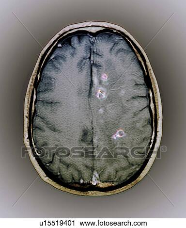
Tapeworm cysts in the brain. Magnetic resonance imaging (MRI) scan of an axial section through the brain of a 25 year old patient showing cysts (neurocysticercosis, purple) from a tapeworm infection. The cysts have been highlighted by the injection of gad Stock Image

Tapeworm cysts in the brain. Magnetic resonance imaging (MRI) scan of an axial section through the brain of a 25 year old patient showing cysts (neurocysticercosis, purple) from a tapeworm infection. The cysts have been highlighted by the injection of gad
u15519401 | Lushpix | Royalty Free
Add to Lightbox
Share Image
Keywords
25-29 years, axial view, biology, brain, brain tissue, cavity, contrast, cross section, cyst, diagnosis, gadolinium, grey background, head, healthcare, human anatomy, human biology, human body part, infection, injected, magnetic resonance imaging, medical, medical scan, medical treatment, medicine, mri, neurocysticercosis, one person, part of the body, pathogen, tapeworm cyst, technology, top view, young adult, stock image, images, royalty free photo, stock photos, stock photograph, stock photographs, picture, pictures, graphic, graphics, fine art prints, print, poster, posters, mural, wall murals, u15519401
25-29 years, axial view, biology, brain, brain tissue, cavity, contrast, cross section, cyst, diagnosis, gadolinium, grey background, head, healthcare, human anatomy, human biology, human body part, infection, injected, magnetic resonance imaging, medical, medical scan, medical treatment, medicine, mri, neurocysticercosis, one person, part of the body, pathogen, tapeworm cyst, technology, top view, young adult, stock image, images, royalty free photo, stock photos, stock photograph, stock photographs, picture, pictures, graphic, graphics, fine art prints, print, poster, posters, mural, wall murals, u15519401
Show Keywords
- Mobile/Small Web Resolution (150 KB)2.9" x 3.4" @ 72dpi JPG13
- Web Resolution (500 KB)5.4" x 6.3" @ 72dpi JPG32
- Low Resolution (1 MB)7.6" x 8.9" @ 72dpi JPG68
- Medium Resolution (10 MB)5.8" x 6.7" @ 300dpi JPG140
- High Resolution (28 MB)9.6" x 11.3" @ 300dpi JPG215
- Ultra High Resolution (50.3 MB)12.9" x 15.1" @ 300dpi JPG250
View larger image sizes
View Lushpix license agreement


