• My Account
  • View Cart
  • Lightboxes
  • English  
    • عربي
    • 中文(简体)
    • Český
    • Dansk
    • Deutsch
    • English
    • Ελληνικά
    • Español
    • Suomi
    • Français
    • עברית
    • हिन्दी
    • Magyar
    • Italiano
    • 日本語
    • 한국어
    • Lietuvių kalba
    • Latina
    • Nederlands
    • Norsk
    • Polski
    • Português
    • Română
    • Slovenčina
    • Svenska
    • Türkçe
    • 中文(繁體)

Stock Photography and Stock Footage

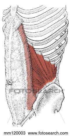
Internal oblique muscle Drawing

Drawing - Internal oblique muscle. Fotosearch

Internal oblique muscle

mm120003 | LifeART | Royalty Free

  • Add to Lightbox

  • Download a Preview

  • Share Image

Keywords
stock illustration, royalty free illustrations, stock clip art icon, stock clipart icons, logo, line art, EPS picture, pictures, graphic, graphics, drawing, drawings, vector image, artwork, EPS vector arts, fine art prints, print, poster, posters, mural, wall murals, mm120003
Show Keywords
  • PX
  • IN
  • CM
Display credit pricing
Display cash pricing
  • Ultra High Resolution (2.3 MB)
    669 x 1203 px JPG
    2.2" x 4" @ 300dpi JPG
    5.7 x 10.2 cm @ 300dpi JPG
    $29.95
    19
View LifeART license agreement
Save up to 80% when you buy with Credits

Best-Selling Related Images


External oblique muscle
Abdominal muscle, artwork
Inferior view of eyeball with musculature.
Superior view of eyeball with musculature.
Horizontal section of inguinal canal showing indirect inguinal hernia.
Headache due to muscle pain, artwork
Posterolateral view of the posterior wall. Latissimus dorsi muscle is partially reflected.
Anterior view of abdominal cavity, skin and musles of anterior abdominal wall cut away to reveal contents undisturbed. Wall section cut from this illustration shown in 2. 18B.
Anterior cross-section view of eye with muscle attachments.
Cross-section of cardiac muscle showing internal anatomy.
Lateral view of eye with muscle attachments.
Stabilization of oblique or spiral fractures by direct fixation with screws.
See All

More Related Images

See All

  • Course of ventral nerve ramus L1 in abdominal wall.
    Course of ventral nerve ramus L1 in abdominal wall.
  • Anterior view of coverings of spermatic cord and testis. Aponeurosis of external oblique muscle incised and reflected, revealing deeper internal oblique muscle Window in internal oblique muscle exposes deeper transversus abdominis muscle.
    Anterior view of coverings of spermatic cord and testis. Aponeurosis of external oblique muscle incised and reflected, revealing deeper internal oblique muscle Window in internal oblique muscle exposes deeper transversus abdominis muscle.
  • Rectum, anal canal and anal sphincter.
    Rectum, anal canal and anal sphincter.
  • Anterior view of the right lower abdominal muscles. The external oblique muscle has been partially resected to reveal the internal oblique and aponeurosis. The conjoint tendon is shown formed by the arching fibers of the internal oblique and the transve
    Anterior view of the right lower abdominal muscles. The external oblique muscle has been partially resected to reveal the internal oblique and aponeurosis. The conjoint tendon is shown formed by the arching fibers of the internal oblique and the transve
  • Posterolateral view of the posterior wall. External oblique muscle is incised and turned laterally, and the internal oblique muscle is incised and turned medially exposing the subcostal (T12) and iliohypogastric nerve (L1). Square in insert indicates or
    Posterolateral view of the posterior wall. External oblique muscle is incised and turned laterally, and the internal oblique muscle is incised and turned medially exposing the subcostal (T12) and iliohypogastric nerve (L1). Square in insert indicates or
  • Posterior view of the abdominal region illustrating the internal abdominal oblique muscle and its bony attachments. Both sides of muscle are shown.
    Posterior view of the abdominal region illustrating the internal abdominal oblique muscle and its bony attachments. Both sides of muscle are shown.
  • Adductor magnus muscle, posterior view.
    Adductor magnus muscle, posterior view.
  • Anterior view of the anterior ends of the inferior intercostal spaces. Portions of muscles have been dissected to reveal intercostal nerves. The image to the right indicates the orientation of 1. 18.
    Anterior view of the anterior ends of the inferior intercostal spaces. Portions of muscles have been dissected to reveal intercostal nerves. The image to the right indicates the orientation of 1. 18.
  • Stabilization of transverse or short oblique and short butterfly fractures by direct fixation with plates and screws.
    Stabilization of transverse or short oblique and short butterfly fractures by direct fixation with plates and screws.
  • Needle penetrating the muscle to gain access to the chest cavity.
    Needle penetrating the muscle to gain access to the chest cavity.
  • Anterior view of the male inguinal region. Internal oblique muscle is reflected, and the spermatic cord retracted. Square in insert image of right pelvis indicates area shown in main image.
    Anterior view of the male inguinal region. Internal oblique muscle is reflected, and the spermatic cord retracted. Square in insert image of right pelvis indicates area shown in main image.
  • Lateral view of coverings of spermatic cord, layers incised and pulled back.
    Lateral view of coverings of spermatic cord, layers incised and pulled back.

Recently Viewed Images

  • Internal oblique muscle
    Internal oblique muscle
 
  • About Us
  • Contact Us
  • Satisfaction Guarantee
  • Website Terms of Use
  • DMCA Procedure
  • Privacy Policy
  • Help
  • CD Store
  • Licensing

Publitek, Inc. dba Fotosearch
Find the Perfect Photo Fast!®

Fotosearch Royalty Free Stock Photography
Digital images licensed by Publitek, Inc.
Fotosearch and Photosearch are trademarks of Fotosearch, LLC
All rights reserved © 2025-09-11