• My Account
  • View Cart
  • Lightboxes
  • English  
    • عربي
    • 中文(简体)
    • Český
    • Dansk
    • Deutsch
    • English
    • Ελληνικά
    • Español
    • Suomi
    • Français
    • עברית
    • हिन्दी
    • Magyar
    • Italiano
    • 日本語
    • 한국어
    • Lietuvių kalba
    • Latina
    • Nederlands
    • Norsk
    • Polski
    • Português
    • Română
    • Slovenčina
    • Svenska
    • Türkçe
    • 中文(繁體)

Stock Photography and Stock Footage

Opponens digiti minimi Stock Illustration
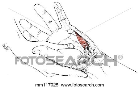
Stock Illustration - Opponens digiti minimi. Fotosearch

Opponens digiti minimi

mm117025 | LifeART | Royalty Free

  • Add to Lightbox

  • Download a Preview

  • Share Image

Keywords
stock illustration, royalty free illustrations, stock clip art icon, stock clipart icons, logo, line art, EPS picture, pictures, graphic, graphics, drawing, drawings, vector image, artwork, EPS vector arts, fine art prints, print, poster, posters, mural, wall murals, mm117025
Show Keywords
  • PX
  • IN
  • CM
Display credit pricing
Display cash pricing
  • Ultra High Resolution (2.4 MB)
    1191 x 705 px JPG
    4" x 2.4" @ 300dpi JPG
    10.1 x 6 cm @ 300dpi JPG
    $29.95
    19
View LifeART license agreement
Save up to 80% when you buy with Credits

Best-Selling Related Images


Abductor digiti minimi
Flexor digiti minimi
Bones of forearm and hand showing muscle attachments, anterior view.
Middle layer of muscles on the posterior forearm.
Anterior view of the deep flexors of the digits and related structures.
Anterior view of the deep palmar arch and adductor pollicis with the digiti minimi and abductor and flexor brevis cut away.
Transverse section through adductor pollicis; for level of section, see insert (line A).
Sole of foot. First layer of muscles and plantar nerves.
Second layer of muscles of sole of foot.
Synovial sheaths on dorsum of wrist.
Posterior view of the extensor digiti minimi muscle with bony attachments.
First layer of muscles of sole of foot, digital nerves and arteries.
See All

More Related Images

See All

  • Motor distribution of the radial nerve to muscles in the posterior compartment of the forearm.
    Motor distribution of the radial nerve to muscles in the posterior compartment of the forearm.
  • Dorsal view of tendons on the dorsum of hand and extensor retinaculum.
    Dorsal view of tendons on the dorsum of hand and extensor retinaculum.
  • Posterior view of the deep structures on extensor aspect of forearm.
    Posterior view of the deep structures on extensor aspect of forearm.
  • Third layer of muscles of sole of foot.
    Third layer of muscles of sole of foot.
  • Anterior view of superficial dissection of the palm with skin, superficial fascia, palmar aponeurosis, and thenar and hypothenar fasciae removed.
    Anterior view of superficial dissection of the palm with skin, superficial fascia, palmar aponeurosis, and thenar and hypothenar fasciae removed.
  • Synovial sheaths and tendons at ankle. A. Tendons at ankle, lateral view. The ankle, subtalar and calcaneocuboid joints are exposed to reveal their positions.
    Synovial sheaths and tendons at ankle. A. Tendons at ankle, lateral view. The ankle, subtalar and calcaneocuboid joints are exposed to reveal their positions.
  • Joints of inversion and eversion. C. Posterior part of foot with talus removed, superolateral view. Note the posterior, middle, and anterior calcaneus facets, the spring ligament, and the facet on the navicular constitute the socket for the talus.
    Joints of inversion and eversion. C. Posterior part of foot with talus removed, superolateral view. Note the posterior, middle, and anterior calcaneus facets, the spring ligament, and the facet on the navicular constitute the socket for the talus.
  • Blood supply of foot. B. Transverse section near bases of metatarsals. The lateral plantar artery courses between the first and second layers of muscles, and then between the third and fourth layers. Green, first layer; orange and gray, second layer; brow
    Blood supply of foot. B. Transverse section near bases of metatarsals. The lateral plantar artery courses between the first and second layers of muscles, and then between the third and fourth layers. Green, first layer; orange and gray, second layer; brow

Recently Viewed Images

  • Opponens digiti minimi
    Opponens digiti minimi
 
  • About Us
  • Contact Us
  • Satisfaction Guarantee
  • Website Terms of Use
  • DMCA Procedure
  • Privacy Policy
  • Help
  • CD Store
  • Licensing

Publitek, Inc. dba Fotosearch
Find the Perfect Photo Fast!®

Fotosearch Royalty Free Stock Photography
Digital images licensed by Publitek, Inc.
Fotosearch and Photosearch are trademarks of Fotosearch, LLC
All rights reserved © 2025-09-11