• My Account
  • View Cart
  • Lightboxes
  • English  
    • عربي
    • 中文(简体)
    • Český
    • Dansk
    • Deutsch
    • English
    • Ελληνικά
    • Español
    • Suomi
    • Français
    • עברית
    • हिन्दी
    • Magyar
    • Italiano
    • 日本語
    • 한국어
    • Lietuvių kalba
    • Latina
    • Nederlands
    • Norsk
    • Polski
    • Português
    • Română
    • Slovenčina
    • Svenska
    • Türkçe
    • 中文(繁體)

Stock Photography and Stock Footage

Mucosa of small intestine, cross section Drawing
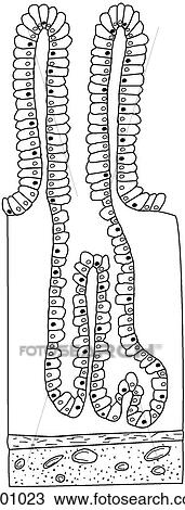
Drawing - Mucosa of small intestine, cross section. Fotosearch

Mucosa of small intestine, cross section

h201023 | LifeART | Royalty Free

  • Add to Lightbox

  • Download a Preview

  • Share Image

Keywords
stock illustration, royalty free illustrations, stock clip art icon, stock clipart icons, logo, line art, EPS picture, pictures, graphic, graphics, drawing, drawings, vector image, artwork, EPS vector arts, fine art prints, print, poster, posters, mural, wall murals, h201023
Show Keywords
  • PX
  • IN
  • CM
Display credit pricing
Display cash pricing
  • Ultra High Resolution (9.6 MB)
    1130 x 2972 px JPG
    3.8" x 9.9" @ 300dpi JPG
    9.6 x 25.2 cm @ 300dpi JPG
    $29.95
    19
View LifeART license agreement
Save up to 80% when you buy with Credits

Best-Selling Related Images


Cross section of the human small intestine, computer illustration.
Cross section of the human small intestine, computer artwork.
Cross section of the human small intestine, computer illustration.
Microscopic cross section view of the small intestine.
Cross section of the human small intestine, computer artwork.
See All

More Related Images

  • Frontal cross-section of the large intestine (colon) showing internal anatomy.
    Frontal cross-section of the large intestine (colon) showing internal anatomy.
  • Cross-section of small synovial joint showing progression of disease of rheumatoid arthritis: step 4.
    Cross-section of small synovial joint showing progression of disease of rheumatoid arthritis: step 4.
  • Cross-section of small synovial joint showing progression of disease of rheumatoid arthritis: step 2.
    Cross-section of small synovial joint showing progression of disease of rheumatoid arthritis: step 2.
  • Cross section of the interior surface of the transverse colon showing the semilunar folds and teniae coli on the smooth-surfaced wall.
    Cross section of the interior surface of the transverse colon showing the semilunar folds and teniae coli on the smooth-surfaced wall.
  • Cross section of the interior surface of the ileum, distal part, showing an absence of plicae circulares. Solitary lymph nodules stud the wall.
    Cross section of the interior surface of the ileum, distal part, showing an absence of plicae circulares. Solitary lymph nodules stud the wall.
  • Orientation drawing of the small intestine shows location of sections.
    Orientation drawing of the small intestine shows location of sections.
  • Deep dissection of abdominal region. Most of small intestine has been removed as well as the mesocolon and peritoneum exposing the underlying psoas muscle and aortic and vena caval bifurcations. Superior gastric plexus is also visible lying on the left
    Deep dissection of abdominal region. Most of small intestine has been removed as well as the mesocolon and peritoneum exposing the underlying psoas muscle and aortic and vena caval bifurcations. Superior gastric plexus is also visible lying on the left

Recently Viewed Images

  • Mucosa of small intestine, cross section
    Mucosa of small intestine, cross section
 
  • About Us
  • Contact Us
  • Satisfaction Guarantee
  • Website Terms of Use
  • DMCA Procedure
  • Privacy Policy
  • Help
  • CD Store
  • Licensing

Publitek, Inc. dba Fotosearch
Find the Perfect Photo Fast!®

Fotosearch Royalty Free Stock Photography
Digital images licensed by Publitek, Inc.
Fotosearch and Photosearch are trademarks of Fotosearch, LLC
All rights reserved © 2025-09-05