• My Account
  • View Cart
  • Lightboxes
  • English  
    • عربي
    • 中文(简体)
    • Český
    • Dansk
    • Deutsch
    • English
    • Ελληνικά
    • Español
    • Suomi
    • Français
    • עברית
    • हिन्दी
    • Magyar
    • Italiano
    • 日本語
    • 한국어
    • Lietuvių kalba
    • Latina
    • Nederlands
    • Norsk
    • Polski
    • Português
    • Română
    • Slovenčina
    • Svenska
    • Türkçe
    • 中文(繁體)

Stock Photography and Stock Footage

Anterior ankle & dorsum of foot Stock Illustration
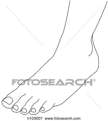
Stock Illustration - Anterior ankle & dorsum of foot. Fotosearch

Anterior ankle & dorsum of foot

h103007 | LifeART | Royalty Free

  • Add to Lightbox

  • Download a Preview

  • Share Image

Keywords
stock illustration, royalty free illustrations, stock clip art icon, stock clipart icons, logo, line art, EPS picture, pictures, graphic, graphics, drawing, drawings, vector image, artwork, EPS vector arts, fine art prints, print, poster, posters, mural, wall murals, h103007
Show Keywords
  • PX
  • IN
  • CM
Display credit pricing
Display cash pricing
  • Ultra High Resolution (7.6 MB)
    1573 x 1690 px JPG
    5.2" x 5.6" @ 300dpi JPG
    13.3 x 14.3 cm @ 300dpi JPG
    $29.95
    19
View LifeART license agreement
Save up to 80% when you buy with Credits

Best-Selling Related Images


Ankle joint and joints of dorsum of foot. B. Distended ankle joint, anterior view.
Anomalous dorsalis pedis artery, lateral view.
Anterior view of the external anatomy of the right leg.
Anterior view of the bones of the right leg.
View of dorsum of foot demonstrating the deep muscles and tendons of that area.
Anterior view of the muscles of the right leg.
View of dorsum of foot demonstrating the muscles and tendons of that area.
Anterior view of the external anatomy of the right leg (ankle and foot).
Medial ankle and foot. B. Foot raised as in walking, medial view.
Anterior view of the muscles of the right leg (tibialis anterior muscle).
Dorsum of foot. B. Dorsalis pedis vein and deep fibular nerve, cut short.
Anterior view of the skeletal anatomy of the right leg (tibia).
See All

More Related Images

See All

  • Dorsum of foot. D. Arterial supply. The dorsal arterial arch is formed by the dorsalis pedis artery, which also contributes to the plantar arch formed by the medial and lateral plantar arteries.
    Dorsum of foot. D. Arterial supply. The dorsal arterial arch is formed by the dorsalis pedis artery, which also contributes to the plantar arch formed by the medial and lateral plantar arteries.
  • Dorsum of foot. C. Bones.
    Dorsum of foot. C. Bones.
  • Anterior view of the muscles of the right leg (adductor muscles of the medial thigh).
    Anterior view of the muscles of the right leg (adductor muscles of the medial thigh).
  • Ligaments of ankle, medial view.
    Ligaments of ankle, medial view.
  • Arteries and nerves of anterior and lateral aspects of leg and dorsum of foot. B. Tibiofibular articulations and their relationships to arteries of the leg, anterior view.
    Arteries and nerves of anterior and lateral aspects of leg and dorsum of foot. B. Tibiofibular articulations and their relationships to arteries of the leg, anterior view.
  • Scheme of motor distribution of the fibular (peroneal) nerve in the anterior and lateral crural compartments and the dorsum of the foot.
    Scheme of motor distribution of the fibular (peroneal) nerve in the anterior and lateral crural compartments and the dorsum of the foot.
  • Bones of leg, showing muscle attachments in anterior and lateral compartments. A. Tibia and fibula, and dorsum of foot, anterior view.
    Bones of leg, showing muscle attachments in anterior and lateral compartments. A. Tibia and fibula, and dorsum of foot, anterior view.
  • Transverse section and MRI through ankle. A. Transverse section. Insert image indicates level of dissection.
    Transverse section and MRI through ankle. A. Transverse section. Insert image indicates level of dissection.
  • Arteries and nerves of anterior and lateral aspects of leg and dorsum of foot. A. dissection, anterolateral view. The anterior crural muscles were removed, and the fibularis (peroneus) longus muscle was excised.
    Arteries and nerves of anterior and lateral aspects of leg and dorsum of foot. A. dissection, anterolateral view. The anterior crural muscles were removed, and the fibularis (peroneus) longus muscle was excised.

Recently Viewed Images

  • Anterior ankle & dorsum of foot
    Anterior ankle & dorsum of foot
 
  • About Us
  • Contact Us
  • Satisfaction Guarantee
  • Website Terms of Use
  • DMCA Procedure
  • Privacy Policy
  • Help
  • CD Store
  • Licensing

Publitek, Inc. dba Fotosearch
Find the Perfect Photo Fast!®

Fotosearch Royalty Free Stock Photography
Digital images licensed by Publitek, Inc.
Fotosearch and Photosearch are trademarks of Fotosearch, LLC
All rights reserved © 2025-09-09