• My Account
  • View Cart
  • Lightboxes
  • English  
    • عربي
    • 中文(简体)
    • Český
    • Dansk
    • Deutsch
    • English
    • Ελληνικά
    • Español
    • Suomi
    • Français
    • עברית
    • हिन्दी
    • Magyar
    • Italiano
    • 日本語
    • 한국어
    • Lietuvių kalba
    • Latina
    • Nederlands
    • Norsk
    • Polski
    • Português
    • Română
    • Slovenčina
    • Svenska
    • Türkçe
    • 中文(繁體)

Stock Photography and Stock Footage

Joint, synovial, equine Stock Illustration
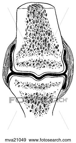
Stock Illustration - Joint, synovial, equine. Fotosearch

Joint, synovial, equine

mva21049 | LifeART | Royalty Free

  • Add to Lightbox

  • Download a Preview

  • Share Image

Keywords
stock illustration, royalty free illustrations, stock clip art icon, stock clipart icons, logo, line art, EPS picture, pictures, graphic, graphics, drawing, drawings, vector image, artwork, EPS vector arts, fine art prints, print, poster, posters, mural, wall murals, mva21049
Show Keywords
  • PX
  • IN
  • CM
Display credit pricing
Display cash pricing
  • Ultra High Resolution (4.2 MB)
    905 x 1624 px JPG
    3" x 5.4" @ 300dpi JPG
    7.7 x 13.7 cm @ 300dpi JPG
    $29.95
    19
View LifeART license agreement
Save up to 80% when you buy with Credits

Best-Selling Related Images


Hock joint, equine
Hock joint, equine
Joint development, equine
Femoropatellar joint, equine
Distal interphalangeal (coffin) joint, equine
Coffin, pastern, and fetlock joint, equine
Lateral view of skull featuring temporomandibular joint.
Cross-section of a synovial joint to show the progression of osteoarthritis (in four stages).
Generic symbol for a synovial joint encased in membrane lined cavity where lubricating fluid is secreted.
Cross-section of a synovial joint to show the progression of osteoarthritis: stage 4.
Cross-section of small synovial joint showing progression of disease of rheumatoid arthritis: step 1.
Cross-section of small synovial joint showing progression of disease of rheumatoid arthritis: step 4.
Cross-section of a synovial joint to show the progression of osteoarthritis: stage 3.
See All

More Related Images

See All

  • Distended knee joint, lateral view.
    Distended knee joint, lateral view.
  • Open knee joint, anterior view.
    Open knee joint, anterior view.
  • Anterior view of the surfaces of radiocarpal (wrist) joint; image on left shows the structures of the hand and wrist superimposed on outline of hand.
    Anterior view of the surfaces of radiocarpal (wrist) joint; image on left shows the structures of the hand and wrist superimposed on outline of hand.
  • Hip joint. B. Posterior view.
    Hip joint. B. Posterior view.
  • Superior view of transverse section of the intervertebral disc and ligaments, with the nucleus pulposus removed and the epiphyseal plate exposed.
    Superior view of transverse section of the intervertebral disc and ligaments, with the nucleus pulposus removed and the epiphyseal plate exposed.
  • Posterior view of the interior of the shoulder joint; joint is exposed by cutting away the posterior part of the capsule and the head of the humerus.
    Posterior view of the interior of the shoulder joint; joint is exposed by cutting away the posterior part of the capsule and the head of the humerus.
  • Transverse section through elbow joint; image to the right shows level of section.
    Transverse section through elbow joint; image to the right shows level of section.
  • Medial aspect of knee. C. Articularis genu muscle. This muscle, deep to the vastus intermedius muscle, consists of a few fibers arising from the anterior surface of the femur and inserting into the synovial capsule of the knee joint, which it retracts dur
    Medial aspect of knee. C. Articularis genu muscle. This muscle, deep to the vastus intermedius muscle, consists of a few fibers arising from the anterior surface of the femur and inserting into the synovial capsule of the knee joint, which it retracts dur
  • Anterior view of the ligaments of distal radioulnar, radiocarpal, and intercarpal joints; hand is shown forcibly extended.
    Anterior view of the ligaments of distal radioulnar, radiocarpal, and intercarpal joints; hand is shown forcibly extended.

Recently Viewed Images

  • Joint, synovial, equine
    Joint, synovial, equine
 
  • About Us
  • Contact Us
  • Satisfaction Guarantee
  • Website Terms of Use
  • DMCA Procedure
  • Privacy Policy
  • Help
  • CD Store
  • Licensing

Publitek, Inc. dba Fotosearch
Find the Perfect Photo Fast!®

Fotosearch Royalty Free Stock Photography
Digital images licensed by Publitek, Inc.
Fotosearch and Photosearch are trademarks of Fotosearch, LLC
All rights reserved © 2025-09-10