Fotosearch Royalty Free Stock Photography
Digital images licensed by Publitek, Inc.
Fotosearch and Photosearch are trademarks of Fotosearch, LLC
All rights reserved © 2026-01-29
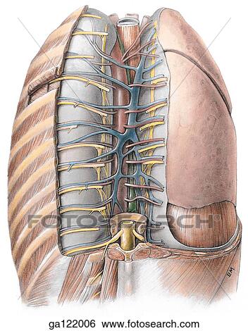
Posterior view of the mediastinum, thoracic vertebral column and thoracic cage are removed on right. On left, ribs and intercostal musculature are removed to angles of ribs. Parietal pleura partially removed on right to reveal visceral pleura covering r Stock Illustration

Posterior view of the mediastinum, thoracic vertebral column and thoracic cage are removed on right. On left, ribs and intercostal musculature are removed to angles of ribs. Parietal pleura partially removed on right to reveal visceral pleura covering r
ga122006 | LifeART | Royalty Free
Add to Lightbox
Share Image
Keywords
a1.72, anatomy, angles, aorta, artery, atlas, azygos, cage, column, communicantes, cord, covering, diaphragm, dissector, dorsal, duct, dural, grant's, hemiazygos, intercostal, left, lobe, lung, lungs, mediastinum, muscle, musculature, nerve, parietal, partially, pleura, posterior, primary, rami, ramus, removed, ribs, right, sac, spinal, splanchnic, sympathetic, thoracic, trunk, vein, vertebral, visceral, stock illustration, royalty free illustrations, stock clip art icon, stock clipart icons, logo, line art, EPS picture, pictures, graphic, graphics, drawing, drawings, vector image, artwork, EPS vector arts, fine art prints, print, poster, posters, mural, wall murals, ga122006
a1.72, anatomy, angles, aorta, artery, atlas, azygos, cage, column, communicantes, cord, covering, diaphragm, dissector, dorsal, duct, dural, grant's, hemiazygos, intercostal, left, lobe, lung, lungs, mediastinum, muscle, musculature, nerve, parietal, partially, pleura, posterior, primary, rami, ramus, removed, ribs, right, sac, spinal, splanchnic, sympathetic, thoracic, trunk, vein, vertebral, visceral, stock illustration, royalty free illustrations, stock clip art icon, stock clipart icons, logo, line art, EPS picture, pictures, graphic, graphics, drawing, drawings, vector image, artwork, EPS vector arts, fine art prints, print, poster, posters, mural, wall murals, ga122006
Show Keywords
- Low Resolution (3.6 MB)13.8" x 17.6" @ 72dpi JPG45
View LifeART license agreement


